See how beautiful the world can look through a microscope
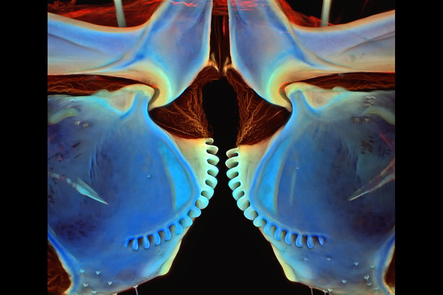
A zebra fish “selfie,” a butterfly’s curly proboscis and the fangs of a centipede. Welcome to the beautiful — and sometimes strange — world of photomicrography.
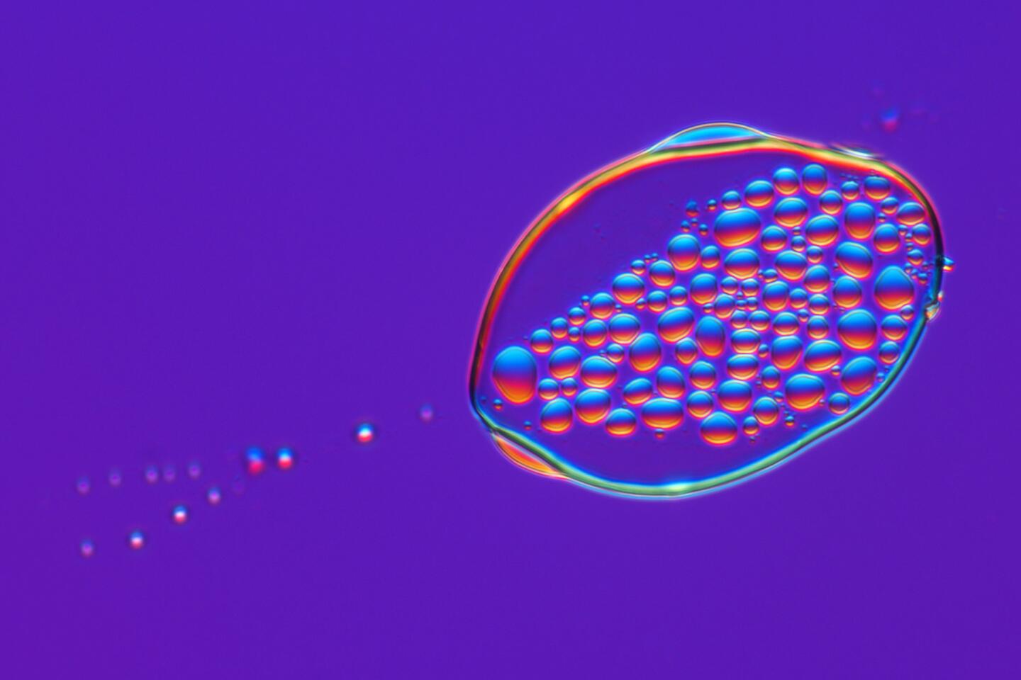
Nikon Instruments has unveiled the winners of its annual Small World Contest. The images celebrate the intersection of art and science, revealing the amazing, minuscule details of the natural world.
The top image was created by Oscar Ruiz, who captured an extreme close-up of a 4-day-old zebra fish embryo. Ruiz studies facial development at the University of Texas MD Anderson Cancer Center, and he hopes zebra fish can help scientists learn how facial abnormalities, such as cleft lip and cleft palate, form in humans.
Ruiz developed innovative techniques for creating time-lapse images of the developing embryo’s face, which he uses to track the movement of facial cells as the fish grows. Knowing this can help pinpoint genetic mutations that give rise to facial deformities.
See the most-read stories in Science this hour »
Other winning images captured magnified images of human neurons, mouse eye cells, intricately arranged microscopic zooplankton and the tiny scales of a butterfly’s wing. Although Nikon’s judges have already spoken, you can still vote for your favorite on the contest’s Facebook page.
Click on the gallery above or scroll down to see some of our favorite images from this year’s contest.
1st Place
Oscar Ruiz
Four-day-old zebra fish embryo
2nd Place
Douglas L. Moore
Polished slab of Teepee Canyon agate
3rd Place
Rebecca Nutbrown
Culture of neurons (stained green) derived from human skin cells and Schwann cells, a second type of brain cell (stained red)
4th Place
Jochen Schroeder
Butterfly proboscis
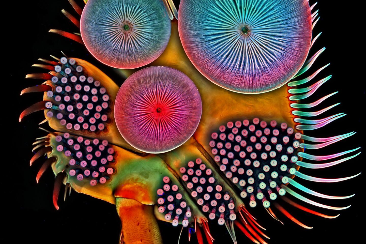
5th Place
Igor Siwanowicz
Front foot of a male diving beetle
7th Place
David Maitland
Leaves of Selaginella (lesser club moss)
11th Place
Francis Sneyers
Scales of a butterfly wing underside (Vanessa atalanta)
13th Place
Walter Piorkowski
Poison fangs of a centipede (Lithobius erythrocephalus)
14th Place
Keunyoung Kim
Mouse retinal ganglion cells
16th Place
Stefano Barone
Sixty-five fossil zooplankton carefully arranged by hand in Victorian style
16th Place
Jose Almodovar
Slime mold
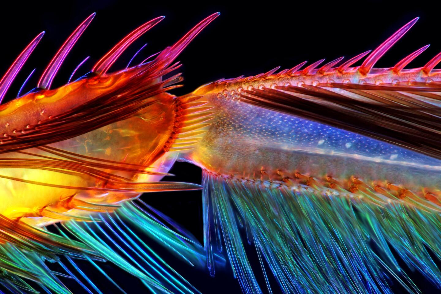
18th Place
Pia Scanlon
Parts of wing-cover (elytron), abdominal segments and hind leg of a broad-shouldered leaf beetle (Oreina cacaliae)
Honorable mentions and images of distinction
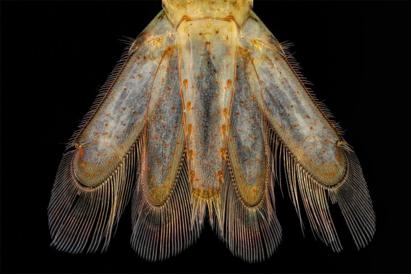
Charles Krebs
Tail of a small shrimp
Jacek Myslowski
Algae
Igor Siwanowicz
Gears coupling the hind legs of a planthopper nymph.
Erno Endre Gergley
Green bottle fly
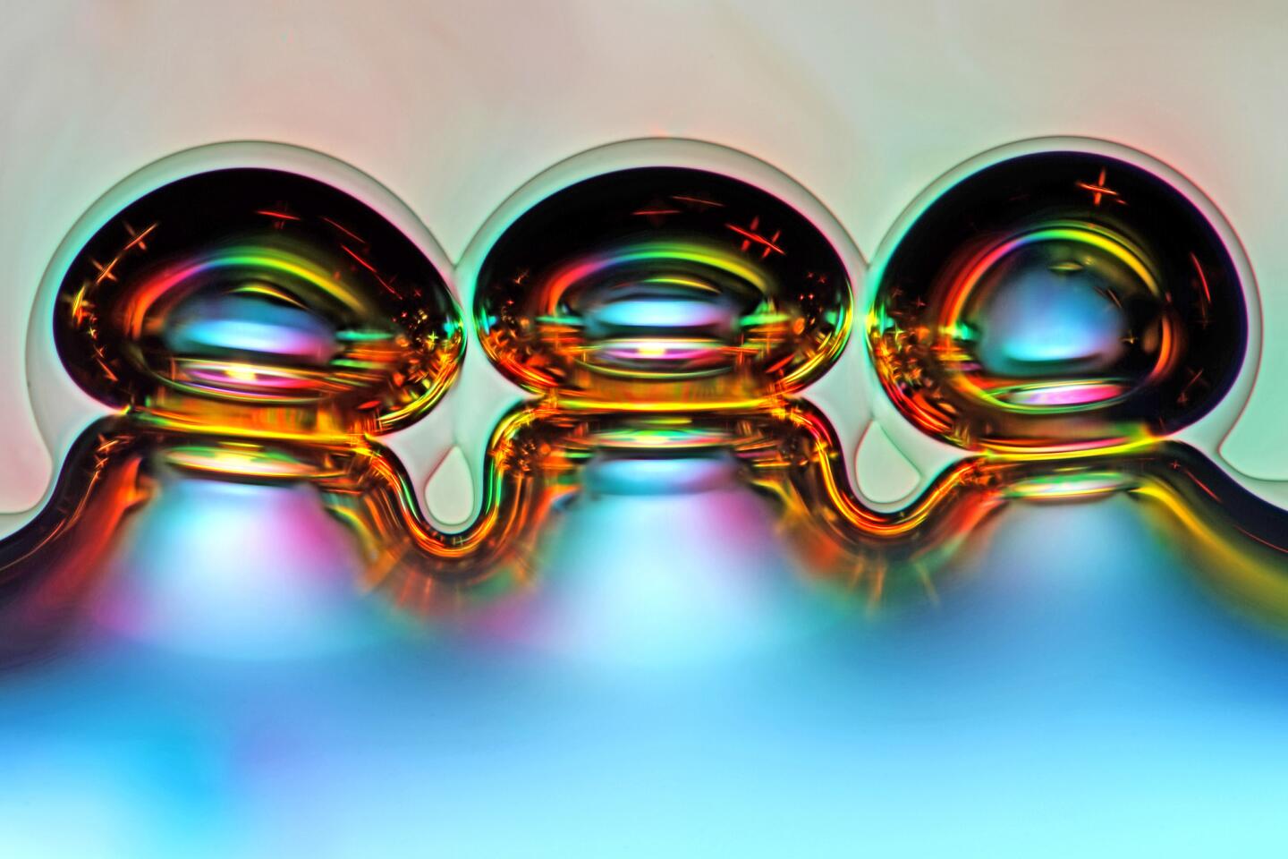
James Hayden
Air bubbles evaporating in tequila
Talley Lambert
Actin (pink), mitochondria (black), and DNA (red) in a bovine pulmonary artery endothelial cell.
Actin (pink), mitochondria (black), and DNA (red) in a bovine pulmonary artery endothelial cell.
Jacek Myslowski
Water mite
Jochen Schroeder
Wasp eyes
Follow me on Twitter seangreene89 and "like" Los Angeles Times Science on Facebook.
MORE SCIENCE NEWS
Science explains why refrigerators sap the flavor from ripe tomatoes
5,000 years ago, rodents were apparently considered food in part of Europe
Surprise! The universe has 10 times as many galaxies as scientists thought
